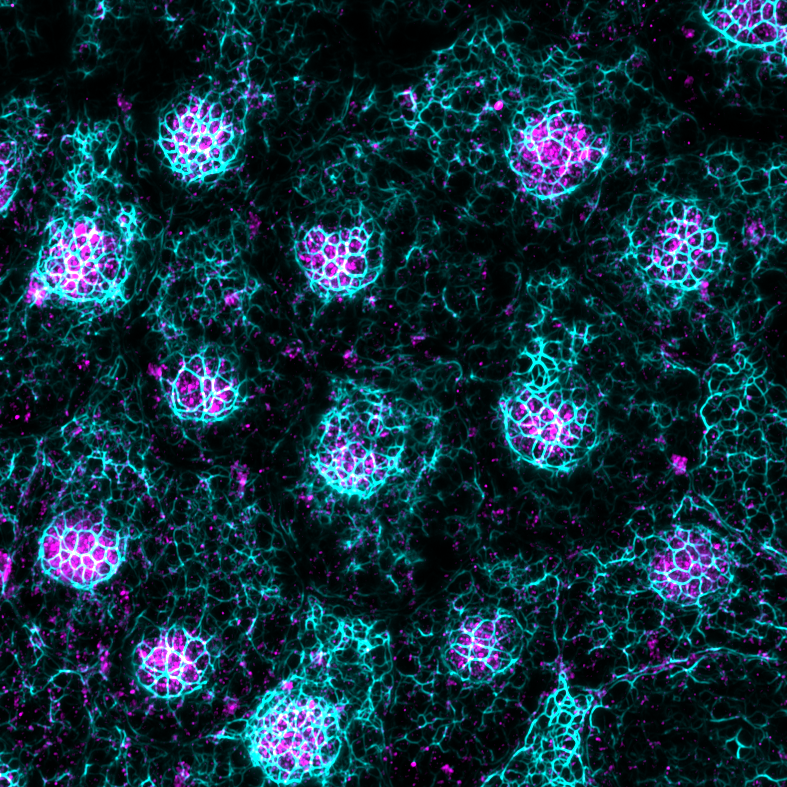
To reproduce the neuroepithelium, we use retinal organoids to study the relation of RE monolayer mechanobiology with photoreceptors cells and their interaction with other cell layers. The figure and video show the three-dimensional cellular organization of the organoids and their differentiation steps.
To study the effect biochemical and physical cues on RE mechanics, we mimic BrM using hydrogels systems of controlled stiffness and functionalization. Hydrogels not only better mimic physiological stiffnesses, but also give us the possibility to employ mechanobiology techniques that allow both qualitative and quantitative evaluation of adhesive forces on the ECM (traction force microscopy) and at cell-cell junctions (monolayer stress microscopy). Different functionalization techniques can be used to study cell adhesion: full leght ECM proteins can be crosslinked on the gel surface or specific ECM-derived peptides can be bound on gold nanoparticles.
Bottom-up investigation of ECM cues in regulating RE mechanobiology
Stem-cell Based Retina Research Model
RE cells are tightly bound to the underlying basement membrane, which is part of a thicker layer of extracellular matrix called Bruch’s membrane. While the basment membrane components carry the biochemical information for RE adhesion, deeper layers of the Bruch's membrane, composed of fibrillar collagen types I, III and V and elastin fibers, are proposed to determine physical properties of RE.
The retina detects light via photoreceptor cells and outer segments (POS), whose homeostasis depends on direct contact with the retinal epithelium (RE). This epithelium tightly adheres to the Bruch’s membrane, which defines its function. Still, the relationship between extracellular matrix biochemistry, physical properties and retinal epithelial mechanobiology has not been addressed. After development, retinal epithelial cells do not proliferate, so the epithelium cannot adapt to the extracellular remodelling that occurs with age (*). This opens the unexplored question of how mechanical forces control the cellular and tissue function of the retina (i.e., retinal mechanobiology) in normal ageing and in age-related macular degeneration.
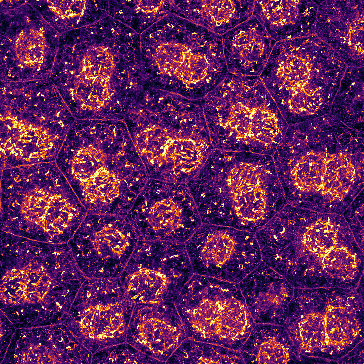
In the laboratory, we use stem-cell derived RE to reproduce in-vitro the retinal epithelium. After few weeks in culture, the cells acquire characteristic features of the in-vivo equivalent such as polarity (Ezrin staining) and apical microvilli.